
Have you ever considered how your own knee muscles function? They are crucial to our daily movements, but do you know what makes up the knee joint and how it functions?
Knee joint anatomy refers to the structure and organization of the knee joint, a hinge joint linking the thigh bone with the shin bone and the patella. Ligaments, tendons, and muscles support the knee joint and include various components such as cartilage, synovial fluid, and menisci. Understanding the knee joint anatomy is important for diagnosing and treating knee injuries and conditions.
The knee is a huge and intricate joint in the human body. It allows us to walk, run, leap, and even sit down because it supports our weight. Understanding the knee joint anatomy is crucial for maintaining health and avoiding injuries.
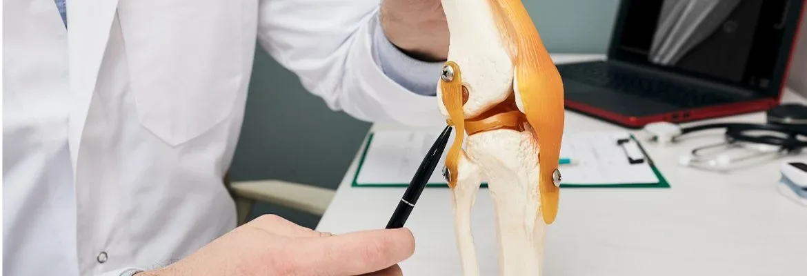
I. Knee Joint Anatomy

Knee Joint Anatomy
One of the most frequently utilized joints in the body is the knee. Its functions include bearing the body’s weight and making movement possible. Knowing the structure of the joint is crucial for keeping it healthy and functioning properly.
Several crucial components make up the anatomy of the knee joint.
- Bones
- Ligaments
- Menisci
- Bursae
- Muscles
· Bones:
The knee has three bones: the femur, tibia, and patella. These bones work together to enable leg movement. They provide flexibility, stability, and strength to the knee. The connection between the femur, patellofemoral joint, and the tibia forms the joint. These bones come into contact during movement, allowing us to bend our knees for different activities.
When issues like arthritis take hold of this complex anatomy, it can cause further damage, such as osteophytes- small growths on bones usually located around joints. These growths can interfere with decreased range of motion due to friction from one bone rubbing against another in areas such as where the femur meets the tibia and where the patella glides down its groove in the patella tendon with leg movements.
· Ligaments:
Those four ligaments are referred to as the collateral ligaments: the anterior cruciate ligament (ACL), the posterior cruciate ligament (PCL), the medial collateral ligament (MCL), and the lateral collateral ligament (LCL), all of them collateral ligaments which operate together to offer stability to lateral surface of the knee joint. These flat collateral ligaments help stabilize joints and prevent excessive movement.
Knee and ligament injuries or ruptures are a common injury that can occur. A ligament is a full band of connective tissue that joins bones or cartilage.
· Menisci:
The medial and lateral menisci are two thin, C-shaped pieces of fibrocartilage in the knee joint between the femur and tibia. These important structures act as shock absorbers, protecting the articular surface of the tibia and assisting in knee rotation. They are incompletely covering the articular surface, providing only partial coverage to allow for movement within the joint. The medial meniscus and lateral meniscus can easily become damaged due to trauma or degeneration. Unfortunately, due to having limited access to a blood supply, these injuries are much more difficult to recover than other parts of the body – yet remain vital components in keeping joints healthy and stable due to their stabilizing function against ligaments
· Bursae:
The knee joint contains several small fluid-filled sacs called bursae. These help to reduce friction between the leg bones, tendons, and ligaments in the joint. The bursae found in the anatomy of the knee joint are a synovial membrane, fluid-filled sacs that act as a protective cushion between moving structures. They are integral for preventing wear and tear on these structures, thus aiding in extending their lifespan. The knee joint has four main bursae: suprapatellar, prepatellar, infrapatellar, medial and lateral semimembranosus.
A suprapatellar bursa is located proximal tibia, above the kneecap patella, and its function is to offer a gliding surface for the quadriceps tendon as it passes over the patella. The prepatellar bursa is a deep pocket located at the top of the knee, between the skin and the patella; its role revolves around protecting this vulnerable section of skin from damage due to the movement of the patella. The infrapatellar bursa provides protection between the bottom part of the patellofemoral joint, the femur bone and the top of the tibia bone, where they form the knee joint capsule itself.
· Muscles:
Several large muscles surround the knee joint, including the quadriceps, hamstrings, and calf muscles. These muscles work together to move the joint and support the body during physical activity.
Understanding the knee joint anatomy is important for preventing injury and maintaining joint health. Proper stretching, strengthening, and conditioning of the muscles surrounding the knee can help avoid damage and minimize potential health problems such as osteoarthritis. If you experience discomfort in your knee joint, getting medical attention to rule out any serious underlying conditions is important.
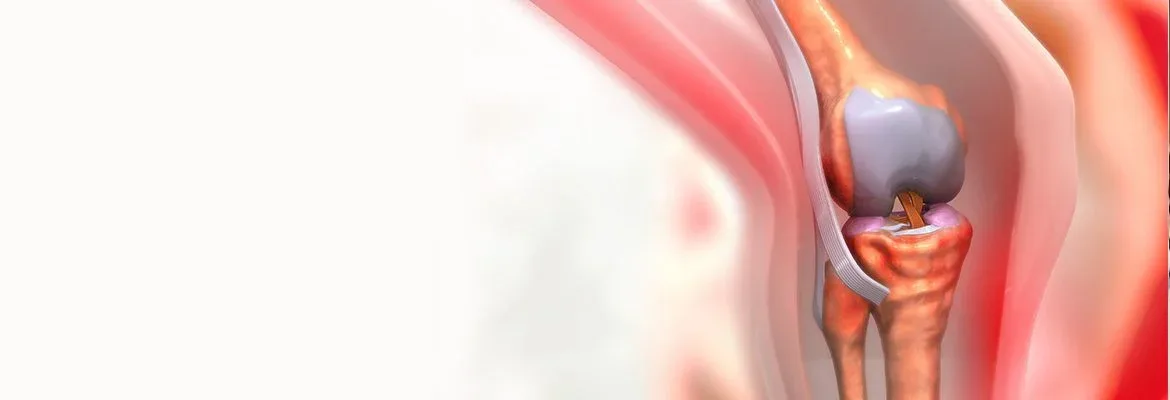
II. Knee Cartilages

Knee Joint Anatomy
Knee cartilage is a crucial component of the knee joint that helps in shock absorption and facilitates smooth movement. Damage or injury to the knee cartilage can cause severe pain and discomfort, decreasing quality of life. In this section, we will discuss knee cartilage and its types.
Knee cartilage is a smooth, rubbery soft tissue covering the bones’ ends in the knee joint. It is a cushion between the knee bones and assists in the smooth gliding of the joint during movement. Knee cartilage is composed of chondrocytes, collagen, and proteoglycans.
· Types of Knee Cartilage
There are two types of knee cartilage:
· Articular Cartilage
Articular cartilage is a specialized type of cartilage covering the ends of bones forming a joint. It has a smooth, white surface designed to withstand the stresses of movement and weight-bearing. The primary purpose of articular cartilage is to minimize friction between the bones during joint movement while evenly distributing weight across the joint to decrease the risk of injury and wear and tear. Articular cartilage comprises chondrocytes, specialized cells that produce and maintain the extracellular matrix that gives the tissue unique properties. Despite its resilience, articular cartilage is vulnerable to damage and degeneration, leading to pain, stiffness, and loss of function in the affected joint.
· Meniscus Cartilage
The meniscus cartilage is the C-shaped connective tissue acting as a shock absorber between the front thigh bone and shinbone. It is composed of fibrocartilage and is located on the exterior and interior edges of the knee joint. The meniscus cartilage helps distribute the body’s weight across the anatomy of the knee joint and prevents the bones from rubbing against each other. The medial meniscus is a crescent-shaped piece of fibrocartilage on the knee joint’s inner side. It functions as an impact absorber and aids in weight distribution across the anatomy of the knee joint. The medial meniscus is susceptible to injury, particularly in athletes who participate in sports requiring twisting or pivoting motions. Common injuries to the medial and lateral meniscus include tears, strains, and degeneration.
Knee cartilage is a crucial component of the knee joint that helps in shock absorption and facilitates smooth movement. Injuries to knee cartilage can cause pain, swelling, and stiffness in the knee joint. Taking care of your knee joint and seeking medical attention if you encounter any knee pain is essential.
III. Functions of Knee Joint
Knee Joint Anatomy
The knee joint is an incredibly important part of the human knee anatomy itself. It is essential to mobility and stability and is key to everyday activities. The knee joint serves several major functions, most notably supporting the body when standing or moving and helping to keep balance. Without our knees, bending, running, walking, jumping, or climbing stairs would be impossible.
The knee joints also enable us to move our legs in many useful directions. We can extend our legs outwards to walk or twist them aside when changing demands in sports like soccer and basketball. Furthermore, our knees provide stability when standing and sitting, absorbing shocks from sudden movements or falls. The large number of muscles working around the kneecap allows for much flexibility and strength compared to other joints, such as the hip joint and shoulders.
· What Type of Joint Is The Knee?
A synovial joint is composed of a fibrous capsule filled with lubricating fluid. This fluid enables the bones to move smoothly and increases their range of motion. Examples of synovial joints in the body include the shoulder, elbow, wrist, hip, ankle, and toes. They are highly mobile joints that allow for movement in multiple directions, like flexion and extension, as well as some rotational movements.
Synovial joints are surrounded by strong ligaments that provide stability for each part of the synovial joint and help it resist stresses placed on it by external forces. Additionally, tendons around the area attach muscles to bones to increase the range of motion achieved when muscle contractions occur to move. The knee contains a wide array of these muscles, ligaments, and tendons which enable it to function optimally while providing support and stability at the same time.
· Impact of Arthritis on the Joint
The impact of knee arthritis on the joint refers to the negative effects that arthritis can have on the complex structure, function, and mobility of a joint. Pain, stiffness, swelling, and a limited mobility range are all symptoms of knee arthritis, which causes inflammation and harm to the joints. The impact of knee arthritis on the largest joint can differ depending on the severity of the state, the type of arthritis, and the location of the affected joint. In some cases, arthritis can cause irreversible damage to the joint, leading to chronic pain and disability.
IV. Common injuries

The knee is one of the body’s multiplex joints and a frequent injury site because of its complexity and prominence. Knee injuries can occur for various reasons, including sports activities, accidents, and aging.
· 1. ACL Tear
One of the primary four ligaments of the knee is the collateral ligament, the anterior cruciate ligament (ACL) which is the collateral ligaments responsible for knee stability during running and jumping. An ACL tear is a collateral ligament, a prevalent knee injury caused by sudden twisting or pivoting. Symptoms of an ACL tear include pain, swelling, and instability in the knee joint.
· 2. Meniscus Tear
The lateral meniscus, is a C-shaped, elastic disc that cushions the knee joint and aids in weight distribution. A meniscus rupture is a knee injury caused by sudden bending or twisting the knee meniscus. Knee joint signs of a torn meniscus include pain, swelling, and rigidity.
· 3. Knee Sprain
A knee sprain is an injury that may occur if the knee is bent or rotated abruptly. A knee sprain happens when one or more meniscofemoral ligaments in the knee joint are stretched or ruptured. A knee sprain is characterized by pain, edema, and instability in the ankle joint.
· 4. Patellar Tendinitis
Patellar tendinitis, or jumper’s knee, is a common injury due to repetitive stress on the patellar tendon. The patellar tendon links the kneecap to the shinbone and is responsible for helping straighten the knee. Symptoms of patellar tendonitis include pain, swelling, and stiffness in the patellar ligament of the knee joint. Treatment for patellar tendinitis may involve rest, physical therapy, and anti-inflammatory medications.
· 5. Osteoarthritis
Osteoarthritis is a common knee injury caused by gradual joint wear and strain. Osteoarthritis occurs when the cartilage tears in the knee mutually break down, causing pain, swelling, and stiffness in the knee joint. Treatment for osteoarthritis may involve rest, physical therapy, and anti-inflammatory medications.
V. Knee Joint Tests
The knee is particularly vulnerable to damage and degeneration since it is a multiplex joint. Several tests can be performed to assess the health of the knee compound joint and diagnose any underlying conditions.
· 1. Lachman’s Test
The Lachman test is used to determine the stability of the anterior cruciate ligament (ACL). In this position, the patient’s knee was bent at a 20-30 degree angle as they rested on their back. The examiner has one hand on the lower leg and the other on the thigh. The examiner then pulls the lower, medial and lateral rotation of leg forward while pushing the lateral and medial rotation of thigh backward. If there is excessive anterior movement lateral rotation of the lower leg, it may indicate a torn ACL.
· 2. McMurray Test
The McMurray test is used to assess for meniscal tears. The patient was positioned on their back with one knee propped up. The examiner grasps and rotates the patient’s foot while applying pressure to the knee. A meniscal tear may be the cause of a clicking or popping noise.
· 3. Varus/Valgus Stress Test
The Varus/Valgus stress test is used to assess for medial collateral ligament or lateral collateral ligament (MCL/LCL) injuries. The patient was positioned on their back with one knee propped up. The examiner applies pressure to the inside (varus) or outside (valgus) of the knee while stabilizing the knee anatomy the thigh. If there is excessive movement, it may indicate an MCL or LCL injury.
· 4. Patellar Apprehension Test
The Patellar Apprehension Test is employed to evaluate patellar instability. The patient was positioned on their back with one knee propped up. The examiner applies pressure to the patella and moves it laterally. If the patient appears apprehensive or experiences pain, it may indicate patellar instability.
· 5. Apley’s Compression Test
The Apley’s Compression test is used to assess cartilage tears. The patient lies on their stomach with their knee bent. The examiner applies downward pressure to the patient’s heel while rotating the foot. If there is pain or clicking, it may indicate a meniscal tear.
VI. Knee Treatments

Knee joint pain is an ailment that affects millions of people worldwide. Various factors, including injury, aging, and arthritis, can cause it. Fortunately, several treatments available can help alleviate knee joint pain and improve mobility. Here are some of the most effective knee common treatments:
· Physical Therapy
Physical therapy is one of the most useful treatments for knee joint pain. A physical therapist can assist you in strengthening the muscles around your knee joint, which can help reduce pain and improve mobility. They can also teach you to exercise at home to alleviate knee joint pain.
· Medications
Medications such as acetaminophen and ibuprofen can assist in relieving knee joint discomfort. If your knee joint pain is severe, your physician may prescribe harsher medications such as opioids or corticosteroids. Before consuming any medication, it is crucial to consult a physician to ensure its safety and efficacy.
· Injections
Knee, knee joint capsule inflammation can be alleviated with injections. Injections of corticosteroids can reduce inflammation and pain in the knee and joint capsule, while hyaluronic acid injections can lubricate the knee and joint capsule and reduce pain. However, the joint capsule capsules are inappropriate for everyone; you should consult your physician before receiving injections.
· Surgery
In some cases, surgery may be important to treat knee pain. There are several common knee injuries and joint surgeries, including arthroscopy, osteotomy, and knee replacement surgery. Knee replacement surgery is typically reserved for severe cases of knee pain where other treatments have failed.
· Lifestyle Changes
Making lifestyle changes can also help alleviate chronic knee pain. Maintaining a healthy weight can help reduce the stress on your knee joint. In contrast, regular exercise can help strengthen the muscles around your knee compound extended knee joint and improve mobility. Additionally, avoiding high-impact activities like running and jumping can help reduce knee joint pain.
VII. FAQS
- What is the knee joint?
The lower leg bone (femur) and the bone that forms the shin bone (tibia) and the kneecap (patella) come together to form the knee joint.
- What are the primary structures of the knee?
The femur, tibia, patella, lateral meniscus,, ligaments, and cartilage make up the primary components of the knee joint.
- What is the function of the knee joint?
The knee joint provides stability and mobility to the lower leg, allowing us to walk, run, jump, and perform other physical activities.
- What are common knee joint injuries?
Common knee joint injuries include ACL tears, meniscus tears, patellar tendonitis, and osteoarthritis.
- How can I keep my knee joint healthy?
To keep your knee joint healthy, it’s important to maintain a healthy weight, exercise regularly, wear proper footwear, and avoid activities that strain the knee joint excessively.